Frequency of Canines’ Impaction: A Radiographic Study Using Orthopantomograms Among a North African Population
DOI:
https://doi.org/10.61838/Keywords:
Teeth, Denture retention, Orthopantomograms, TunisiaAbstract
Background. Canines represent the keystone of dentition since they maintain the harmony of occlusion and play a major role in dentofacial esthetics. These functions may be altered by several etiologies such as canines’ impaction (CI).
Objective. To investigate the frequency and patterns of CI in a Tunisian population.
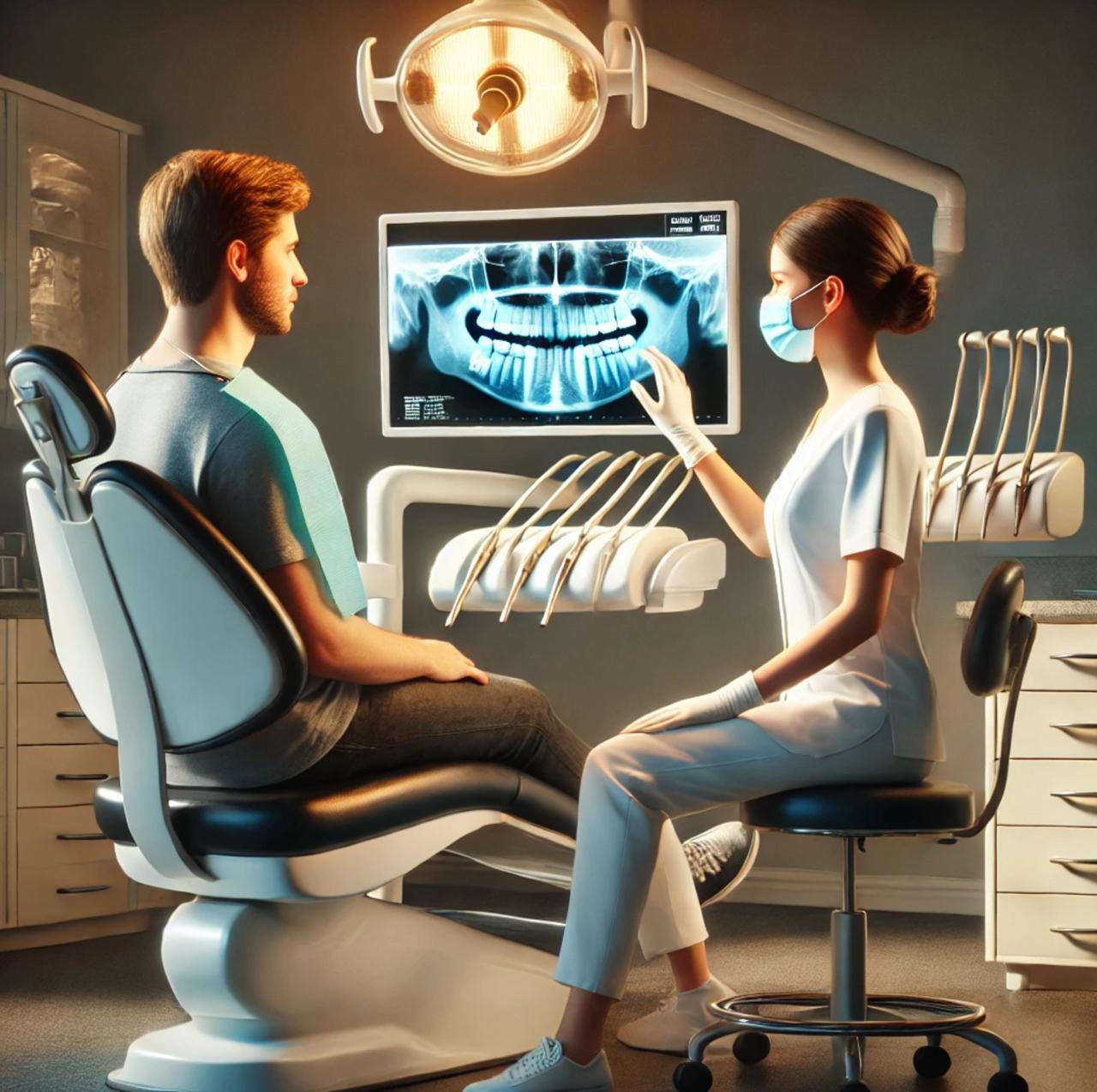
Methods. This was a descriptive retrospective study conducted in the Dental Medicine Department (Fattouma Bourguiba Hospital of Monastir, Tunisia). Patients with a minimum age of 14 years, consulting between January 2013 and December 2022, were included. Orthopantomograms were retrieved for all the consultants. Impacted maxillary canines were classified in terms of tooth direction and position using a validated classification system. Pathologies associated with impacted canines were recorded.
Results. Among the 6462 orthopantomograms analyzed, 136 patients (2.1%) presented CI with no significant difference between males (31.6%) and females (68.4%) (p=0.08). The number of impacted canines was 167 with a statistical difference between arches (73% and 10.5% in the maxilla and the mandible, respectively) (p<0.01), but no significant difference between the left and the right sides (p=0.84). The most common number of impacted canines was one (79.4%). The most common type of impacted maxillary canines was type IV and II (33.5%). One patient was diagnosed with a follicular cyst associated and 12 cases of transmigrated upper impacted canines were found.
Conclusion. Impacted maxillary canine is one of the most common and confusing issues that a dentist can face in his daily practice. Early radiographic examination and diagnosis are essential to identify CI.

Downloads
Published
Issue
Section
License
Copyright (c) 2024 Soumaya Zaalouni, Sinda Yacoub, Wala Guesmi, Salwa Ben Menaa, Nouha Dammak, Mohamed Ben Khelifa (Author)

This work is licensed under a Creative Commons Attribution-NonCommercial 4.0 International License.


